Computerized Thermographic Imaging and Live Blood Analysis Post C19 Injection
This is a lot of conjecture. Beware of misinformation.
I just got this message:
From Ana Maria Mihalcea,
Dear Colleagues,
I interviewed a Brazilian Biologist and Researcher, Felipe Reitz, who is doing Computerized Thermographic Imaging in the C19 injected and correlating this with Live Blood Analysis. The clots are everywhere, even in young athletes in their 20’s, it is absolutely heartbreakingly shocking. These are completely asymptomatic. For the love of humanity, please look at these images and the video we did.
As an Internal Medicine Physician of over 20 years’ experience, I have never seen anything so devastating in its implications. He has found the same thing in children, as young as 2 years old.
If this visual proof does not help doctors stop the jabs, or people not to get them, I do not know what will. Maybe whole-body Computerized Thermography needs to be standard of care for all C19 injected people.
Here is the link to the story which has a link the 1 hour video. I’ve also emailed Reitz.
Pretty much every person I’ve talked to has very serious doubts that the methodology described can show anything useful.
One of my experts wrote:
He is arbitrarily talking about cell size. Talking about PMNs that are not T-cells. He is all over the map. This is the type of presentation that lacks enough credibility to watch all of it. If what you (these people) present up front is garbage, I won’t waste a full hour of my life watching the rest.
Moral of the story: tread very carefully in new territory.
This does not appear to be credible. I’m posting this here to point out that there is lot of information which looks technical but is unreliable.


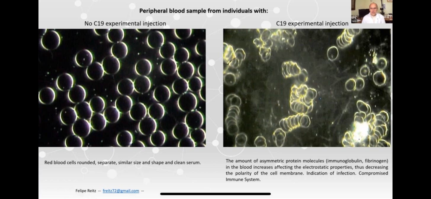
Steve, I am a hematologist and have published extensively on why red cells do what they do. The pictures are poor, but the second picture shows mostly what is commonly called RBC Rouleaux -- stacks of red cells that occur (with or without covid or spikeshots) usually when there is hyperglobulinemia of any cause in the blood. The usual causes are immunogobulins (created in response to any inflammation) or hematologic malignancies that secrete immunoglobulins (like multiple myeloma). Fibrinogen (a key component of blood clotting) is also elevated in inflammation and can cause rouleaux formation. Rouleaux are not an unusual finding although they are not "normal" for most people. (They are relatively common in horse blood, incidentally, because the protein mix in the blood is different.)
There may also be some agglutination in that picture -- hard to tell because bunches of rouleaux can also look like agglutinates. Agglutinates are just clumps of red cells (rather than rolls) due to antibodies that stick to red cells -- because the red cell is nothing but a unit membrane filled with hemoglobin solution, these are not common. Agglutinates are often artifacts of collection (EDTA, an anticoagulant, can cause this) or may indicate immune mediated hemolytic anemia -- but I have not seen that as a covid complication of significance.
I am not selling anyone a machine (as opposed to the picture supplier -- nothing wrong with selling something, but disclosure matters) but have looked at tens of thousands of blood smears and have seen many, many with rouleaux that look just like this for decades. Having said that, I have maintained since 2020 that covid is in large part a hematologic, not respiratory, disease for a host of reasons. And if this hematologic damage is spike-related, than it will be impacted by spikeshots as well. But this picture/explanation does not contribute to thinking about that in my opinion.
Dear Steve. I am a medical doctor who does conventional and "alternative' medicine. I used to do dark field microscopy and am familiar with this. Frankly, i would shy away from these kinds of claims. what you are seeing on the one side with all the clumping of the red blood cells is a phenomena called "rouleaux" stacking. I think it is because of electrostatic charges on the RBC membrane. At any rate, this is not clot formation, as clotting involves a complex interaction with fibrin and platelets, and this is not that. the other important thing to understand that in anyone's dark field blood analysis, you can practically find this rouleaux stacking somewhere on the slide if you hunt for it. Of course, some blood samples are blatantly full of rouleaux and others will be 95 or 96% free of it, but you can almost find it in everyone's sample. so for people to show dark field slides as a sign of some Covid spike or gene-jab spike, is not a good thing to rely on. (BTW, I'm an unjabbed physician who saw the nefariousness of the geng-jab from the get go, so I'm on your side and appreciate all the data analysis and talking points that you bring up regarding the Covid jabs and all vaccines. everything should be questioned -- after all, that is the scientific process!) ron manzanero, md Arlington, Tx